Description
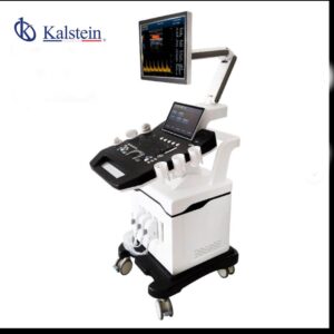
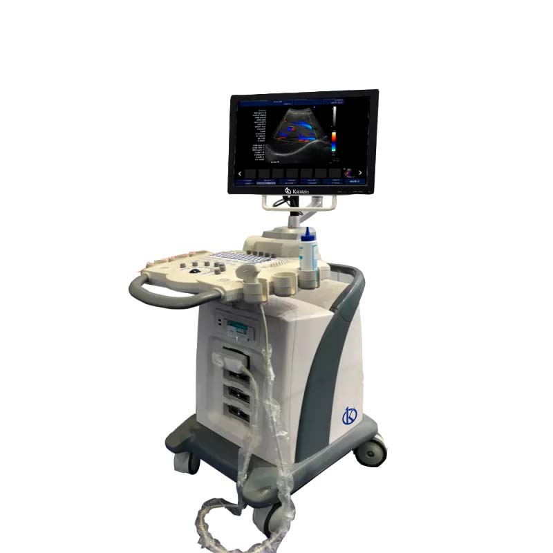
The Color Doppler Ultrasound Cart with Wheels 3D Model YR05149 is a cutting-edge diagnostic tool engineered for precision and ease of use in medical imaging. It boasts a 15-inch LED screen with multilingual functions and supports a variety of imaging technologies such as CF+B mode, PDI, DPDI, TDI, and TSI. Capable of trapezoidal imaging, virtual convex array, and matrix expansion, this ultrasound cart is highly versatile, meeting diverse diagnostic needs. Additional features include automated IMT measurement, endometrial evaluation, and quality control, making it invaluable in fields like obstetrics and cardiology. The integration of the DICOM 3.0 protocol ensures smooth data sharing and printing, while the inclusion of workstations and databases streamlines workflow. Its wheeled, portable design is particularly suited for dynamic healthcare settings. Enhance your practice with this all-encompassing diagnostic solution and generate a customized quote via Kalstein Plus today.
Market Price
Understanding the market pricing for ultrasound equipment is crucial when considering an investment in the Color Doppler Ultrasound Cart with Wheels 3D Model YR05149. While specific prices may vary, it is widely recognized for its advanced technology and versatility, representing excellent value for medical facilities looking to enhance their diagnostic capabilities.
Frequently Asked Questions
How many transducer ports are available? This model offers 4 transducer ports, facilitating simultaneous connections for varied diagnostic tasks.
Can the ultrasound cart connect to external devices? Yes, with multiple connectivity options including USB, Ethernet, VGA, and HDMI ports, it ensures broad compatibility with external devices.
Advantages and Disadvantages
Advantages: This ultrasound cart is exceptionally versatile and portable, featuring advanced imaging methods and user-friendly attributes that simplify a wide array of diagnostic applications.
Disadvantages: The initial investment can be significant for smaller clinics, although the long-term advantages and improvements in efficiency often offset these costs.
Product Use in the Field
This ultrasound cart is utilized predominantly in clinical environments such as obstetrics and cardiology, where precise and adaptable imaging solutions are essential. It allows for swift transitions between imaging modes and supports comprehensive diagnostic assessments, thereby enhancing patient care quality.
Recommendations
It is recommended to perform regular maintenance checks and timely software updates to maximize the longevity and performance of this ultrasound cart. Ensuring that operators receive proper training will enable them to effectively utilize all functions and improve diagnostic results.
Features
- 15″ LED screen with multilingual function
- Model CF+B simultaneous imaging, PDI, DPDI, TDI, TSI support
- Automatic IMT and endometrial measurements
- Seamless DICOM 3.0 protocol integration
- Efficient workflow with integrated workstations and databases
- User-friendly online guidance and control
Technical Specifications
| Model | YR05149 | |||
| Connectivity/Media/Peripherals | ||||
| Transducer Ports | 4 | |||
| USB ports | 4 | |||
| HDD | 64GB (SSD), 120G/200GB SSD (optional) | |||
| foot switch | USB | |||
| Ethernet port | 2(100Mb/1000Mb) | |||
| External screen | VGA,HDMI, | |||
| Printer (Optional) | USB Printer, Digital Laser Printer, B/W Digital Thermal Printer | |||
| Printing area | Image, report, Image+report | |||
| Cine/Picture Memory | ||||
| Memory Cinema | 1200 frames (max) | |||
| Film Review Speed | 1, 2, 4, 8 | |||
| Cinema Review Loop | YES | |||
| Capture function | YES | |||
| DICOM connectivity | DICOM3.0 Compliant | |||
| 3Dsoftware | Built-in 3D software | |||
| Image Storage | Storage Format: PNG, AVI, BMP, JPEG, DICOM Export Video Format: AVI Export Image Format: PNG, JPEG, BMP, DICOM USB Flash Drive |
|||
| Technology | ||||
| Panoramic Imaging Technology Full Digital Signal Processing Technology Multi-Beamforming Technology Spot Reduction Technology Tissue Harmonic Imaging Technology Dynamic Tissue Optimization Technology Duplex and Triplex Synchronous Display Directional Power Doppler Imaging Parameters Imaging Parameters special tissue presets PW Auto Trace Update online CF+B mode on one screen Complex model imaging Automatic IMT measurements Virtual Convex Array Trapezoidal imaging |
||||
| Overall Performance | ||||
| Digital Broadband | 12288 channels | |||
| Beam Former | reprogrammable | |||
| Transmission voltage | Adjustable (15 steps) | |||
| Beamformer Frequency Range | 1~40MHz | |||
| Led monitor | ||||
| Size (diagonal) | fifteen” | |||
| Contrast Ratio | 800: 01: 00 | |||
| Resolution | 1024×768 pixels | |||
| Brightness | 230 cd / m2 | |||
| Color Depth | 24 bit | |||
| Rotation Angle | ±90° | |||
| Gray levels | 256 | |||
| Imaging performance | ||||
| Start Time (max.) | Average < 90 seconds | |||
| Preset Switching Time | Average < 1 second | |||
| Storage Time (Image to disk) | Average < 0.5 seconds | |||
| transducers | ||||
| Research | Convex array probe | Linear Array Probe | intracavitary probe | microconvex probe |
| Frequency | Center 3.5MHz | Central 7.5MHz | Center 6.5MHz | Center 4.0MHz |
| (2.0MHz to 10.0MHz) | (6.0MHz to 10.0MHz) | (5.0MHz to 9.0MHz) | (2.0MHz to 5.5MHz) | |
| field of play | 0.516mm | 0.352mm | 0.216mm | |
| Radio | 60mm | N/A | 10mm | |